– a responsible task, which is both an excellent cross-section of knowledge on a certain subject, as well as an excellent incentive for independence, the manifestation of scientific thinking and information analysis. Of course, when faced with such a serious test for the first time, every student wants to write a laboratory work in biology properly. How to do this will be discussed in this article.
In any case, manuals and manuals will come to the aid of the student, from which one can glean enough information. In addition to studying the theoretical part, the student must also have an understanding of the goals of the workshops and the rules for working with the necessary instruments that will be used during the research.
As a rule, each student must purchase an A4 notebook in which he will make notes about the goals of the work, his actions, the knowledge applied and the instruments used, as well as the results obtained. This notebook is called a laboratory notebook. It is from this that the notes will be compiled; it is the journal that largely answers the question of how to do laboratory work in biology and any other discipline. The main difference between such work and similar research in chemistry is the less stringent safety precautions.
Officially registered work consists of several components:
- Title page.
- Introductory part.
- Practical section.
- Conclusions
And now in more detail about how to do a biology lab:
- The cover page contains information such as information about educational institution, the name of the department, the topic of the work, the name of the student who completed it, as well as the name of the person accepting the work. At the end of the sheet the year and city are usually indicated.
- The introductory part contains the purpose and objectives, as well as general information.
- The practical section reflects the actions of the author, that is, it presents full speed work with all calculations.
- In conclusion there is a very important area of all the work done - the conclusion.
How to write a conclusion for a laboratory work in biology
In the final part, the student presents the results of his practical research. This modest but most important part should not be drawn out, since a meaningful and concise conclusion is the best option. The teacher evaluates this part very carefully. It is important to understand that the conclusion is based on the purpose of the work.
Some tips on how to write a conclusion:
- Try to express your thoughts scientifically.
- Do not pour water.
- Report results and actions taken.
Thus, based on the material in this article, you can get an idea of how to write a laboratory work in biology in order to get a satisfactory grade and consolidate the acquired knowledge.
Conclusions on laboratory work- briefly formulated results of processing measurement results - should be given in the section “Results of measurement processing and conclusions” of the abstract for each laboratory task. The outputs should display the following information:
what was measured and by what method;
what graphs were built;
what results were obtained.
Also, the conclusions should contain a discussion of the constructed graphs and the results obtained: whether the appearance of the experimental graphs coincides or not with theoretical predictions and whether the experimental results coincide or not with the theory. The recommended form for presenting conclusions based on graphs and responses is given below.
|
OUTPUT according to GRAPH (template): The experimentally obtained dependence graph name of the function in words from argument name has the form of a straight line (parabola, hyperbola, smooth curve) and qualitatively coincides with the theoretical dependence of these characteristics, which has the form formula(if the type of dependence is unknown, then it is not necessary to provide it). |
|
OUTPUT based on ANSWER (template): The experimentally obtained value of the quantity full name of the physical characteristic, equal symbol = (average ± error) ·10 degree unit(δ = ___%), within the limits of error coincides (does not coincide) with the tabular (theoretical) value of this value, equal to number, unit of measurement. |
Graphing
1. Graphs are made in pencil on graph paper or on a squared sheet of at least ½ notebook size.
2. A rectangular coordinate system is used with UNIFORM axle markings. Argument values are plotted along the X axis, function values are plotted along the Y axis.
3. The scale and origin are chosen so that the experimental points are located over the entire area of the figure.
4. The scale unit must be a multiple of 1×10 n, 2×10 n 3×10 n etc., where n= …-2, -1, 0, 1, 2, ….
5. Next to the axis the letter designation, order and dimension of the physical quantity are given.
6. Below the graph – the full name of the graph IN WORDS.
7. No lines or marks can be drawn to explain the construction of points on the graph.
Examples:
|
RIGHT |
WRONG |
Title page design
TO
 Report
for laboratory work No.
«__________________________________________________________ __________________________________________________________»
Completed Art. groups
____________________________
Teacher (academic level, title)
____________________________

EXAMPLE OF FORMATING A LABORATORY WORK REPORT
State Autonomous Educational Institution of the Astrakhan Region of Higher Professional Education
"Astrakhan Engineering and Construction Institute"
TODepartment of Physics and Mathematics, Information Technologies
 Report
for laboratory work No. 1.2.
"STUDYING ERRORS IN ACCELERATION MEASUREMENT
FREE FALL USING A MATHEMATICAL PENDULUM"
(name of laboratory work)
Completed Art. ASG groups – 11-10
Ivanov Ivan Ivanovich
Teacher: Ph.D.-M.Sc., Associate Professor.
_____Petrov Sergey Ivanovich

1.09.11 Petrov
1.09.11 Petrov
5.09.11 Petrov
Goal of the work: 1) study of the oscillations of a mathematical pendulum: measuring the period of its oscillations and determining the acceleration of gravity;
2) assessment of random and instrumental measurement errors; studying the dependence of the width of the confidence interval on the number of experiments and confidence probability.
Experimental setup diagram

1 – tripod;
2 – thread lengthl;
3 – load;
4 – stopwatch;
5 cm tape
Calculation formulas
 ,
,
 ;
;
g – acceleration of gravity;
l – thread length;
N – number of oscillations during time t.
Result of thread length measurement: l= 70.5 cm = 0.705 m.
Calculation of constant C
C = (2 5) 2 0,705 = 695,807 696 (m).
Exercise 1. ERROR ASSESSMENT
RESULT 25 MEASUREMENTS
Table 1
|
Experiment number | ||||
Laboratory work No. 1
Variety of plant divisions.
Target: study the diversity of plant divisions.
Lesson objectives:
introduce students to systematics - the science of the diversity and classification of organisms;
reveal the tasks and significance of taxonomy.
During the classes:
I.
Updating knowledge
Filling out the “Kingdoms of Wildlife” diagram.
II.
Learning new material
1.
Expand students' knowledge about the diversity of organisms inhabiting the Earth (teacher's story with elements of conversation).
2.
Introduce students to the concept of “systematics”. Species is the initial unit in taxonomy (teacher's story).
3.
C. Linnaeus is the founder of taxonomy. Double Latin names of species (teacher's story with demonstration of plant and animal species on living objects, herbarium materials, collections).
4.
Modern system organic world. Basic systematic units (categories): species, genus, family, order (order), class, department (type), kingdom.
5.
The meaning of taxonomy.
Laboratory work No. 2
Environmental groups land plants in relation to water
Work plan:
1. Read the description of the ecological groups of plants.
2. Determine which ecological group the plant given to you belongs to.
3. Name the signs of adaptation to the environment of this plant.
4. Give examples of plants found in the Republic of Adygea that belong to this ecological group.
Ecological groups of plants.
Hydatophytes- This aquatic plants, completely or almost entirely immersed in water (elodea, pondweed, water buttercups, duckweed). Once taken out of the water, they quickly die.
The leaves of hydatophytes are thin, often dissected; Variation of leaves (heterophylly) is often expressed. The root system is greatly reduced or absent altogether. Absorption of water and mineral salts occurs over the entire surface of the body. Pollination occurs above water (less often in water), and fruit ripening occurs under water, since flowering shoots carry flowers above water and, after pollination, submerge again.
Hygrophytes - land plants, growing in conditions high humidity air and often on wet soils.
Shade hygrophytes- these are plants of the lower tiers of damp forests (impatiens, thistle, many tropical herbs). Their leaves are most often thin and shady. High water content in the tissues of these plants (80% or more). They die even during a short and mild drought.
Light hygrophytes- these are plants of open habitats, growing on constantly moist soils and in humid air (papyrus, rice, hearts, marsh bedstraw, sundew).
Mesophytes - can tolerate short and not very severe drought. They grow with average moisture, moderately warm conditions and a good supply of mineral nutrition. This is the largest and most heterogeneous group in its composition. This includes trees, shrubs and grasses of various zones, many weeds and most cultivated plants.
Xerophytes- grow in places with insufficient moisture. They are able to regulate water metabolism, so they remain active during short droughts. These are plants of deserts, steppes, sand dunes and dry, highly heated slopes.
Xerophytes are divided into two main types: succulents and sclerophytes.
Succulents- succulent plants with highly developed water-storing parenchyma in different organs: stem plants (cacti, cactus-like euphorbia); leafy (aloe, agave); root (oxalis).
Sclerophytes - externally dry, often with narrow and small leaves, sometimes rolled into a tube. Sclerophytes can be divided into two groups: euxerophytes and stypaxerophytes.
Euxerophytes- these are many steppe plants with rosette, semi-rosette, heavily pubescent shoots (shrubs, some cereals, cold wormwood, edelweiss edelweiss).
Stypaxerophytes- these are narrow-leaved turf grasses (feather grass, thin-legged grass, fescue), the leaves of which are rolled into a tube and have a moist chamber inside.
Laboratory work No. 3
Device of magnifying devices.
Goal of the work: learn how to properly use optical instruments (magnifying glass, light microscope); preparation method.
Equipment and materials: microscope, magnifying glass.
Progress:
Examine a hand-held magnifying glass. What parts does it have? What is their significance?
Study the structure of the microscope. Find the tube, eyepiece, lens, tripod with stage, mirror, screws.
Familiarize yourself with the rules of using a microscope.

2. Adjustment screws
4. Lens
5. Stage
7. Mirror
Laboratory work No. 4
Preparation of a micropreparation of onion scale skin
Purpose: to study the structure of a plant cell.
Equipment: hand magnifying glass, microscope, pipette, glass slide, bandage; part of an onion
PROGRESS.
1. Prepare a preparation of onion skin. To do this, use tweezers to separate the bottom surface of the onion scales and remove the transparent skin.
2. Place the preparation on a glass slide. Examine under a microscope.
3. Examine the cell at high magnification.
4. Draw the structure of a cell in your notebook and label its parts. 
5. Draw a conclusion.
Conclusion: A cell is an integral biological system. The cell is the basic structural unit of a living organism.
Laboratory work No. 5
Composition of plant cells
Target: study the composition of plant cells.
Equipment: bulb, microscope, slide and cover glass, dissecting needle, textbook
Progress:
Prepare glass slide, wipe it with gauze.
Apply 1-2 drops of water on glass.
Dissecting needle remove the skin from inner surface onion scales.
Put a piece of peel into a drop of water and straighten it with the tip of a needle.
Cover peel with a cover glass.
Consider prepared preparation under a microscope.
Sketch in your notebook and label: cell, cell wall, cytoplasm, nucleus.
Sketch diagram of the structure of a plant cell and label: nucleus, cell wall, cytoplasm, chloroplasts, vacuole.

Conclusion: A cell is the simplest structural unit of a living organism. The green color of the plant is given by chlorophyll in the composition of chloroplasts.
Laboratory work No. 6
Cell structure of an Elodea leaf
Target: study the structure of the Elodea leaf cell.
Equipment: Elodea leaf, microscope, slide and cover glass, dissecting needle, textbook.
Progress:
Prepare a microslide of an elodea leaf.
■ Place an Elodea leaf in a drop of water on a glass slide, straighten it with a dissecting needle and cover with a coverslip.
■ Examine the preparation under a microscope. Pay attention to the shape and color of the cells. There are nuclei in living Elodea cells, but usually they cannot be seen.

Conclusion. Nuclei and chlorophyll grains are clearly visible in the cells (at higher magnification). The lower layer of smaller cells is clearly visible, intercellular spaces and the outlines of the cells of the upper layer are visible.
Laboratory work No. 7
The structure of an animal cell.
Goal: compare the structure of plant and animal cells and find out what their similarities indicate.
A cell is the main structural, functional and reproductive element of a living organism, its elementary biological system. Depending on the structure and set of cell organelles, all organisms are divided into kingdoms - prokaryotes and eukaryotes. Plant and animal cells are classified in the kingdom of eukaryotes. They have a number of similarities and differences.
General signs:
1) membrane structure of organelles;
2) the presence of a formed nucleus containing a chromosome set;
3) a similar set of organelles, characteristic of all eukaryotes;
4) similarity in the chemical composition of cells;
5) the similarity of the processes of indirect cell division (mitosis);
6) similarity of functional properties (protein biosynthesis), use of energy conversion;
7) participation in the process of reproduction.
Conclusion: the similarity in the structural and functional organization of plant and animal cells indicates their common origin and their belonging to eukaryotes. Their differences are related to in different ways nutrition: plants are autotrophs, and animals are heterotrophs.
Laboratory work No. 8
The structure of the integumentary and synthesizing tissue of plants
Target: get acquainted with the types of tissues of a plant organism, the features of their structure in connection with the function they perform.
Equipment: micropreparations “Longitudinal section of a corn stalk”, “Cross section of a pumpkin root”, “Root structure”; microscopes; tables “Cellular structure of the root”, “Root and its zones”, “Internal structure of the leaf”.
Instruction card1. Consider the microslide “Root Structure” (Fig. 1). Find educational fabric. Us. 30 of the textbook, read about the location of educational tissue, the features of its structure in connection with the function it performs. Enter the data into the table.
 Rice. 1. Internal structure of the root: 1 – root cap (integumentary tissue) protects the zone of dividing cells; 2 – zone of dividing cells (educational tissue) carries out root growth in length
 2. Examine the root cap. Determine the type of tissue that forms it. Us. 30 of the textbook read about this type of fabric. Enter the data into the table.
Table. Plant tissues
| Type of fabric | Location | Structural features | Functions |
| Educational | |||
| Pokrovnaya | |||
| Mechanical | |||
| Conductive | |||
| Main |
3. On the micropreparation “Longitudinal section of a corn stalk”, examine the mechanical tissue of the stalk. Please note that the cells of this tissue have thickened, lignified membranes, and there is no living content. Read about this fabric on p. 30 textbook. Enter the data into the table.
4. Look at the drawing of conductive tissue in the textbook on p. 31. Compare it with what you saw under the microscope (Fig. 2), read the information about this tissue. Enter the data into the table.

Rice. 2. Conductive tissues of the stem: 1 – sieve tubes of the bast (conducting organic matter from leaves to all organs); 2 – vessels of wood (carrying minerals dissolved in water from the root to all organs)
5. To study the main leaf tissue, consider microslides prepared by the teacher (Fig. 3, 4). This is a thin cross section of a Tradescantia leaf. Pay attention to the structural feature of this tissue - the presence of chloroplasts, which contain the pigment chlorophyll. It gives plants green color. Read about the function of this fabric on p. 31 textbooks. Enter the data into the table.

Rice. 3. Internal structure of the leaf: 1 – leaf skin (leaf protection, cover); 2 – basic tissue (photosynthesis, cells contain chloroplasts); 3 – conductive bundle (conduction of substances, strengthening of veins, mechanical tissue); 4 – stomata (water evaporation, gas exchange)

Rice. 4. Leaf peel. 1 – leaf skin (integumentary tissue): cells fit tightly to each other, protecting the leaf from damage
6. Draw a conclusion about the presence of tissues, their different structures and answer the questions:
– How is the structure of tissue related to the function it performs?
– Why do the cells of the integumentary tissue adhere closely to each other?
– How to distinguish the main tissue from the cover tissue?
Laboratory work No. 9
The structure of connective tissues of animals.
Target:
Equipment: microslides “Epithelial tissue”, “Loose connective tissue”, microscopes, table “Structure diagram of an animal cell”.
Progress:


Rice. 1. Types of animal tissues:
A – epithelial tissue; I – loose connective tissue
1. Consider the microslide “Epithelial tissue” (Fig. 1, A). Find the epithelial cells, pay attention to the features of their structure (the cells fit tightly to each other, there is no intercellular substance). Draw the drug. Look at the picture and read necessary information. Enter the data into the table.
2. Examine the microslide “Loose connective tissue” (Fig. 1, AND). Pay attention to the structural features of the fabric (presence large quantity intercellular substance). Draw the drug.
3. Fill out the table.
| Fabric name | Location | Structural features | Functions performed |
| Connective A) bone B) cartilaginous | Dense intercellular substance loose intercellular substance | 1. Support 2. Support and protection |
|
| B) fat | Fat layers | 3. Protective |
|
| Blood vessels | liquid intercellular substance. General: Cells are spaced apart; there is a lot of intercellular substance. | 4. Transport |
Conclusion: Connective tissue consists of a basic substance - cells and intercellular substance - collagen, elastic and reticular fibers. It performs supporting, protective and nutritional (trophic) functions.
Laboratory work No. 10
The structure of muscle and nervous tissues of animals.
Target: get acquainted with the tissues of the animal body, the features of their structure depending on the function performed.
Equipment: "Smooth muscle", "Nervous tissue", microscopes, table "Structure diagram of an animal cell".
Progress:
1. Examine the microslide “Muscle tissue” (Fig. B). Pay attention to the structural features of muscle cells (these are spindle-shaped mononuclear cells). Draw the drug. Look at the picture, read the information about the types, properties of muscle tissue and its function. Enter the data into the table.
2. Examine the microslide “Nervous tissue” (Fig. D). Pay attention to the structural features nerve cells(consist of a body and numerous processes of two types). Draw the drug. Look at the picture, read the information about the properties of nervous tissue and its function. Enter the data into the table.
| Fabric name | Structure | Functions | Examples |
| Muscular | Smooth muscle, consists of elongated cells with rod-shaped nuclei. Cross-striped muscle tissue consists of long, multinucleated fibers with clearly visible transverse striations. | gives shape to the body, supports, protects internal organs. | Movement of animals, ability to respond to irritation (amoeba). |
| Cells (neurons) are stellate in shape with long and short processes | perceives irritation and transmits excitation to muscles, skin, other tissues, organs; ensure coordinated functioning of the body. | forms the nervous system, is part of nerve ganglia, spinal cord and brain. |


Conclusion: Nervous - to lettuces (neurons) are stellate in shape with long and short processes. The function transmits stimulation to muscles, skin, and other tissues. Muscle gives shape to the body, supports and protects internal organs.
Practical work № 1
The influence of light on the growth and development of plants.
Tasks:
Observe the progress of seed germination and plant development in different conditions.
Apply the results obtained in biology lessons and in life.
Growth and nutrition of the seedling. The cells of the root, stem and bud of the embryo, feeding, divide, grow, and the embryo turns into a seedling. When a seed germinates, a root appears first. As it develops, it outstrips other organs of the embryo, quickly strengthens in the soil and begins to absorb water and minerals from it.

Until the seedling reaches the soil surface, organic substances stored in the seed are used for its growth and development. But if they run out before photosynthesis begins, the seedling may die. Therefore, to increase the productivity of cultivated plants great importance has strict adherence to the timing and rules of sowing.
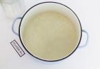
"The influence of light on plant development."
Radish sprouts were created different conditions. Some were grown in the light, others in the dark. The photo shows that the plants, placed in a dark place, began to lag behind in development, became weak, turned yellow, and then completely died. The picture shows before and after the experiment.


From my experience, I concluded that plants develop well only in the light.
Conclusion: For seed germination, the following conditions are necessary: heat, air and water. And for normal height and the development of plants after germination also requires light.
Practical work No. 2
Similarities and differences between plant, animal and fungal cells.
Target: study the similarities and differences between plant, animal and fungal cells.
All three main groups of organisms are
animals,
plants
They are eukaryotes. However, the structure of their cells is not the same. These differences, along with feeding habits, formed the basis for dividing the eukaryotic superkingdom into three kingdoms.
animal cell does not have a dense cell wall. It lacks vacuoles characteristic of plants and some fungi. The polysaccharide glycogen usually accumulates as a reserve energy substance.
Majority plant and fungal cells, like prokaryotic cells, it is surrounded by a hard cell membrane, or wall. However, their chemical composition is different. While the base of the wall plant cell is a polysaccharide cellulose, mushroom the cell is surrounded by a wall, largely consisting of the nitrogen-containing polymer chitin.
Plant cells always contain plastids, while animals and mushrooms no plastids. Reserve substance for most plants the polysaccharide starch serves, and the bulk mushrooms, like animals,- glycogen.
Handout
Laboratory work in biology.
Laboratory work No. 1.
Topic: “Composition of seeds.”
Look at the table on the board. Name the parts of a seed. Conclude why, by studying the composition of seeds, we can judge the chemical composition of the plant.
1. Following safety rules, light the alcohol lamp and warm up the test tube with the seeds. Place a glass slide near the opening of the test tube. What are you observing?
2. Continuing heating, look at what changes occur in the seeds (color, smell). Draw a conclusion.
3. Using personal experience, guess what will happen next. Stop heating, close the alcohol lamp, and place the test tube in a rack. On your own or using the textbook text (p. 10), make a diagram in your notebook “ Chemical composition cells." Check your notebooks and compare them with the table on the board.
Laboratory work No. 2.
Topic: “Determination of the physical properties of proteins, fats and carbohydrates (starch, sugar).
1. Add water to a small amount of wheat flour and make a ball of dough. How has the dough changed?
2. Wrap a lump of dough in gauze, put it in a glass of water and rinse it. How did the water in the glass change?
3. Drop 1-2 drops of iodine solution into a glass with clean water. How did the color of the water change?
4. Drop 1-2 iodine into a glass of water into which the dough was dipped. How did the color of the contents of the glass change? What can be concluded?
5. Place a sunflower seed between two sheets of white paper; Press down firmly on the seed with the blunt end of a pencil. What happened to the paper? What can be concluded?
6. Discuss what physical properties organic substances can be studied and in what way. Enter the obtained data into the table.
Laboratory work No. 3.
Topic: “Cell structure.”
1. Read the first two paragraphs on p. 16 of the textbook and make a diagram of life forms in your notebook. Give brief description each group and give examples of representatives.
2. Set up the microscope and prepare a preparation of onion skin. Make a drawing in your notebook. Name the clearly visible parts of the cell.
3. Knowing the functions of nucleic acids, think about what role the nucleus can play in a cell?
4. Think about why there is a single set of chromosomes in germ cells, and a double set in body cells? What happens if the set of chromosomes changes?
Laboratory work No. 4.
Topic: “Plant tissues.”
1. Think about whether all cells in a multicellular organism are the same in structure. Justify your answer.
2. Find in the textbook on p. 30 definition of tissue, write down the types of plant tissues in your notebook.
3. Examine the finished tissue micropreparations, make the necessary sketches, formulate a conclusion about the connection between the structure of cells and the function performed.
4. Answer the question: are all cells in a multicellular plant organism the same in structure?
Laboratory work No. 5.
Topic: “Animal tissues.”
1. Using the textbook, p. 32-34, write down the types of animal tissues.
2. Examine microscopic specimens of these tissues.
3. Draw a conclusion about the relationship between the structure and function of cells.
4. Are animal tissues different from plant tissues? Why?
Laboratory work No. 6.
Topic: “Structure of root systems.”
1. Consider the root systems of rye and beans.
2. Find adventitious and lateral roots in the rye root system. Is it possible to find the main root in it?
3. What is the name of the root system of rye? Draw and label its parts.
4. Locate the taproot in the bean root system.
5. Sketch root system beans. Label its parts. What is this type of root system called?
Laboratory work No. 7.
Topic: “Microscopic structure of the root.”
1. Examine the “Cellular structure of the root tip” preparation through a microscope, compare with the figure on p. 42 textbooks, highlight the root zones, name their functions.
2. Using your practical experience, name the functions of the root. Write it down in your notebook.
Laboratory work No. 8.
Topic: “Structure and location of the kidneys.”
1. Consider the herbariums and plants offered to you. What kind of buds do you see? How are they located? Make a drawing.
2. Find small elongated and rounded buds on the shoot. Make a drawing.
3. Using a dissecting knife, make a longitudinal incision into the rounded bud. Using a magnifying glass and dissecting needles, examine its internal structure. What is this kidney called? Make a drawing.
4. Using a dissecting knife, cut lengthwise into the smaller elongated bud. Using a magnifying glass and dissecting needles, examine its structure. What is this kidney called? Make a drawing.
5. Why is a bud called an embryonic shoot?
Laboratory work No. 9.
Topic: “Simple and complex leaves.”
1. Look carefully at the leaves offered to you, divide them into groups and explain on what basis you classified them. Justify your answer.
2. Draw a petiole, sessile, compound leaf in your notebook. Label the drawings.
3. Examine the arrangement of leaves on a plant or herbarium specimen. Compare with the location of the kidneys. Draw a conclusion.
Draw a petiolate, sessile one in your notebook. am leaves, divide them into groups and explain on what basis you classified them
Laboratory work No. 10.
Topic: “Structure of a flower.”
1. Examine the flower, holding it by the peduncle. Pay attention to its size, color, smell, number of parts, think about its significance for the life of the plant.
2. Carefully separate the perianth on a piece of paper.
3. Select the main parts of the flower: stamens, pistil. Consider how they are arranged.
4. Write on a piece of paper the names of the parts of the flower and arrange them according to the names (you can use the textbook text on p. 40).
5. Draw a diagram of the structure of a flower in a notebook and sign it. Draw a conclusion about the role of a flower in the life of a plant.
Laboratory work No. 11.
Topic: “Dry and juicy fruits”
1. Using personal experience and the textbook text (p. 40, second paragraph from the bottom), talk about methods of plant pollination. What happens to a flower after pollination? How is the fruit formed?
3. Fill out the table, give examples of fruits and plants in which they are found, draw a conclusion about the importance of fruits in the life of plants.
Variety of fruits.
Laboratory work No. 12.
Topic: “Structure of seeds of dicotyledonous and monocotyledonous plants.”
1. Review and describe appearance bean seeds. Make a drawing.
2. Using a paring knife, remove the seed coat. What is its role for the seed?
3. Consider the structure of the embryo. Make a drawing and label its main parts.
4. Examine and describe the appearance of a wheat grain. Make a drawing.
5. Using a dissecting needle, try to remove the cover of the caryopsis.
6. Using the textbook drawing and ready-made drug“A grain of wheat. Longitudinal section”, which you can examine with a dissecting loupe, make a drawing “Structure of a wheat grain”; label its main parts.
7. Compare the structure of a bean seed and a grain of wheat. Find similarities and differences.
8. Fill out the diagram:
Laboratory work No. 14.
Topic: “Movement of solutions along the stem”
1. Compare the movement of substances along a tree trunk with their movement along a potato stem (textbook, pp. 74 and 75). Make a movement diagram in your notebook.
2. Examine the microslide “Vascular-fibrous bundle of a linden trunk”, compare with the textbook drawings on p. 74 and 75, cut from a potato tuber. Draw the vascular-fibrous bundles in a notebook and label the drawing.
Laboratory work No. 15.
Topic: “Structure of frog and human blood cells.”
1. Look at microscopic specimens of human and frog blood, compare their sizes and make a drawing in your notebook. Compare with the picture in the textbook.
2. Draw conclusions about what you saw.
Laboratory work No. 16.
Topic: “Bone structure.”
1. Consider the animal bones provided. Determine whose bones these are and what they are called. Divide them into groups according to size and structure.
2. Using the picture in the textbook, name the parts of the bone, make a drawing in your notebook “Structure of bone”, and label it.
Laboratory work No. 17.
Topic: “Movement of the ciliate slipper and earthworm.”
1. Use a pipette to drop a drop of the prepared slipper culture onto a glass slide.
2. Cover the drop with a cover slip. Excess water select using filter paper.
3. Examine the preparation under a microscope (objective 20, eyepiece 15).
4. Observe the beating of the eyelashes.
5. Sketch the appearance of the ciliate.
6. Sketch and describe the stages of movement of an earthworm.